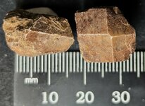
Relative to the specimen listed above, a good friend very familar with this type of mineralization and the general area, suggested to KT that it may be from the Columbia Mine, Marion, Crittenden County, Kentucky. KT investigated the mine and the area using Mindat.com and found a picture by Alan Goldstein, a knowledgeable authority on the mineralization of that region from the Columbia Mine that matches the general look of this specimen, but does not have barite associated with it. So KT has changed the locality information on the pictures to reflect this locality, not the general area. For those interested, His Majesty figured you should have this new info.

You are using an out of date browser. It may not display this or other websites correctly.
You should upgrade or use an alternative browser.
You should upgrade or use an alternative browser.
UV Lights and Fluorescent Minerals - a fun side hobby to metal detecting !
- Thread starter GKL
- Start date
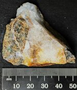
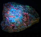
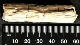
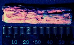
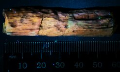
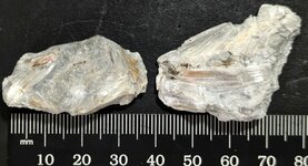
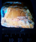
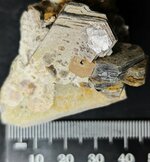
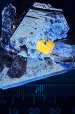
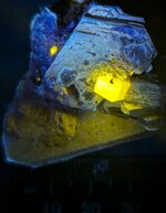
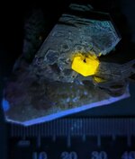
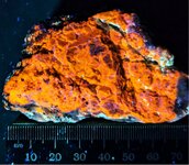
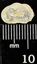
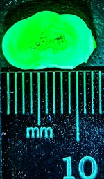
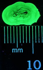
One of the rarer minerals KT obtained for the Royal Collection…..the mineral is Carpathite from the 4th of July Mine, Flint Group, Picacho Peak, San Benito County, California. The Type locality is Russian site and the original name was Karpathite. The mineral is most unusual as it does not fit the typical definition of a mineral….it is organic, chemical formula C24H12, and corresponds to 7 fused benzene rings. A few sites are located in the same region of Picacho Peak for this mineral. These are sites that have been investigated for mercury, so cinnabar often accompanies the Carpathite and Quartz, but not in this specimen! In fact it is a somewhat atypical example, the mineral being encased in the Quartz vein instead of on the surface of the specimen.
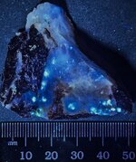
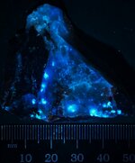
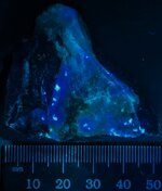
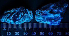
The first picture is in natural light and basically the sample just looks like a normal section of a milky quartz vein with a piece of hostrock attached. The second picture is in LW 365nm and the Carpathite displays as bright blue white spots scattered in the massive milky Quartz vein. The third picture is in MW 310nm and the Carpathite is still nicely responsive in blue white. The fourth picture is in SW254nm and again the Carpathite responds in a blue white, tho not so brightly.
Enjoy the pictures!
Attachments
-
 Carpathite in Quartz vein, 4th of July Mine, Flint Group, Picacho Peak, San Benito Co., CA, na...jpg157.4 KB · Views: 232
 -
 Carpathite in Quartz vein, 4th of July Mine, Flint Group, Picacho Peak, San Benito Co., CA, LW...jpg156.2 KB · Views: 232
 -
 Carpathite in Quartz vein, 4th of July Mine, Flint Group, Picacho Peak, San Benito Co., CA, MW...jpg81.1 KB · Views: 214
 -
 Carpathite in Quartz vein, 4th of July Mine, Flint Group, Picacho Peak, San Benito Co., CA, SW...jpg82 KB · Views: 218
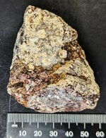
Another arrival from Tuesday!
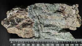
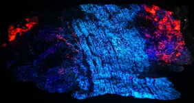
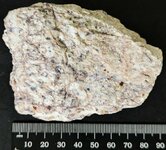
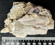
Microcline, also called Orthoclase Perthite by some authors, from the Sterling Hill Mine, Ogdensberg, Sussex County, New Jersey, associated with Calcite. The texture by some has been suggested to be due to metamorphic processes, but it certainly looks like injected fault gouge (mylonized) to KT.
The first picture is in natural light and displays the feldspar zone well at nearly 3 inches across, with highly broken up rock to the right and left in the picture. The specimen is only fluorescent in SW 254nm (the second picture) and the Microcline is a fairly strong whitish blue, and the associated calcite is its typical red orange color.
Enjoy the pictures!
Attachments
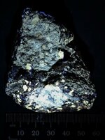
The 3rd and final fluorescent specimen that arrived in the Royal Castle's Mailbox last Tuesday!
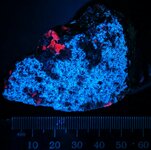
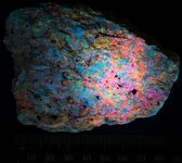
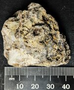
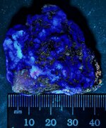
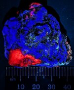
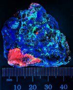
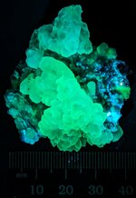
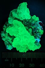
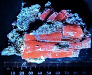
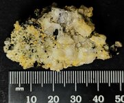
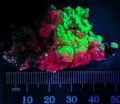
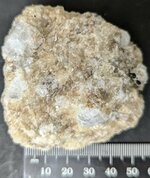
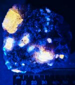
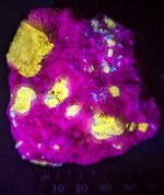
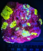
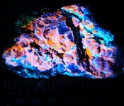
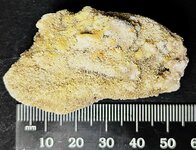
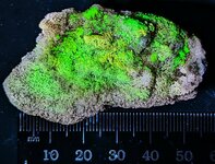
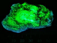
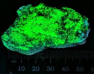
This specimen displays a weathered surface coating of Hydrozincite on a matrix of Calcite and Franklinite (NF, TL) from the Franklin Mine dumps, Franklin, Sussex County, New Jersey. Hydrozincite is a secondary mineral that forms from the weathering of sphalerite or other zinc-bearing minerals and has been well documented as forming on/within the mine dumps, as well as in the oxidized portions of the ore zone within the mines.
The first picture is in natural light and shows an encrustation of white to cream Hydrozincite. The second picture is in LW 365nm with the Hydrozincite responding with a nice blue color. The third picture is in MW 310nm and again the Hydrozincite is blue, and a bit of calcite is giving an orange red response. The fourth picture is in SW 254nm with the Hydrozincite a strong blue and the underlying calcite giving a strong orange red response. Careful examination of the photo also reveals tiny green specks of willemite associated with the calcite!
Enjoy the pictures!
Attachments
-
 Hydrozincite coating Calcite & Franklinite (NF), Franklin Mine dumps, Franklin, Sussex Co., NJ...jpg212.2 KB · Views: 209
 -
 Hydrozincite coating Calcite & Franklinite (NF), Franklin Mine dumps, Franklin, Sussex Co., NJ...jpg174.7 KB · Views: 214
 -
 Hydrozincite coating Calcite & Franklinite (NF), Franklin Mine dumps, Franklin, Sussex Co., NJ...jpg156.1 KB · Views: 208
 -
 Hydrozincite coating Calcite & Franklinite (NF), Franklin Mine dumps, Franklin, Sussex Co., NJ...jpg169 KB · Views: 217
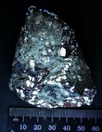
Another specimen from New Jersey!
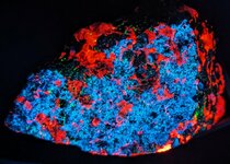
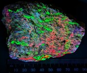
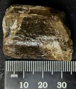
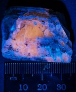
This specimen arrived today and was labeled as Sphalerite, but when KT examined the piece under LW, MW, and SW UV lamps, He had to add both Willemite and Calcite (veins) to the Royal Label. Locality is given as Sterling Hill Mine, Ogdensberg, Sussex County, New Jersey.
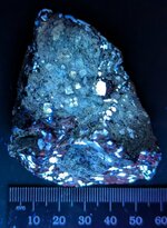
His Majesty took two sets of pictures of this specimen, 8 total pictures, because the fluorescence was so different on one side versus the other. The first set of images are of what KT called the top, and the second set is of the bottom.
First picture is in natural light, and KT is reminded of Ed O’Dells quip that most fluorescent minerals are just cream to gray rocks! HA HA Yes, that is what this looks like, a cream colored rock. The second picture is in LW 365nm, and the Sphalerite gives off a nice orange response..I do not know what the blue is unless it is simply a reflection of the LW light. At first KT thought it might be Hydrozincite as a secondary mineral, but Hydrozincite is most often only responsive to SW UV. The third picture is in MW 310nm, and the Sphalerite has a weaker orange response, while the blue is still there. Note that the size of the specimen precludes me from being able to cover the entire piece with MW UV, so only a round area has strong response. The fourth picture is in SW 254nm, and the Willemite and some spotty Calcite give their typical green and orange-red response, while the Sphalerite is unresponsive.
Now to the reverse side of the specimen. The fifth picture is in natural light. The sixth picture is in LW 365nm and the Sphalerite is again responsive in orange and the unknown mineral displaying blue. The seventh picture is in MW 310nm, and again only an area of the specimen is covered, but the Sphalerite is its typical orange. The eighth and final picture is in SW 254nm, and the Willemite displays green, while previously unnoticed veins of Calcite are a strong red color.
A very interesting specimen, indeed! With this specimen KT also received a two page update on the status of the mines and Mineral Museum which His Majesty put in his old copy of the Fluorescent Minerals of Sterling Hill and Franklin Mines…..and that was very much appreciated!
Thanks!
This specimen arrived today and was labeled as Sphalerite, but when KT examined the piece under LW, MW, and SW UV lamps, He had to add both Willemite and Calcite (veins) to the Royal Label. Locality is given as Sterling Hill Mine, Ogdensberg, Sussex County, New Jersey.
His Majesty took two sets of pictures of this specimen, 8 total pictures, because the fluorescence was so different on one side versus the other. The first set of images are of what KT called the top, and the second set is of the bottom.
First picture is in natural light, and KT is reminded of Ed O’Dells quip that most fluorescent minerals are just cream to gray rocks! HA HA Yes, that is what this looks like, a cream colored rock. The second picture is in LW 365nm, and the Sphalerite gives off a nice orange response..I do not know what the blue is unless it is simply a reflection of the LW light. At first KT thought it might be Hydrozincite as a secondary mineral, but Hydrozincite is most often only responsive to SW UV. The third picture is in MW 310nm, and the Sphalerite has a weaker orange response, while the blue is still there. Note that the size of the specimen precludes me from being able to cover the entire piece with MW UV, so only a round area has strong response. The fourth picture is in SW 254nm, and the Willemite and some spotty Calcite give their typical green and orange-red response, while the Sphalerite is unresponsive.
Now to the reverse side of the specimen. The fifth picture is in natural light. The sixth picture is in LW 365nm and the Sphalerite is again responsive in orange and the unknown mineral displaying blue. The seventh picture is in MW 310nm, and again only an area of the specimen is covered, but the Sphalerite is its typical orange. The eighth and final picture is in SW 254nm, and the Willemite displays green, while previously unnoticed veins of Calcite are a strong red color.
A very interesting specimen, indeed! With this specimen KT also received a two page update on the status of the mines and Mineral Museum which His Majesty put in his old copy of the Fluorescent Minerals of Sterling Hill and Franklin Mines…..and that was very much appreciated!
Thanks!
Attachments
-
 Sphalerite,Willemite, and calcite(veins) top, Sterling Hill Mine, Ogdensberg, Sussex Co., New ...jpg205.7 KB · Views: 224
 -
 Sphalerite,Willemite, and calcite(veins), top, Sterling Hill Mine, Ogdensberg, Sussex Co., New...jpg219.4 KB · Views: 214
 -
 Sphalerite,Willemite, and calcite(veins), top, Sterling Hill Mine, Ogdensberg, Sussex Co., New...jpg165.4 KB · Views: 222
 -
 Sphalerite,Willemite, and calcite(veins), top, Sterling Hill Mine, Ogdensberg, Sussex Co., New...jpg220 KB · Views: 204
 -
 Sphalerite,Willemite, and calcite(veins) bottom, Sterling Hill Mine, Ogdensberg, Sussex Co., N...jpg194.9 KB · Views: 223
 -
 Sphalerite,Willemite, and calcite(veins) bottom, Sterling Hill Mine, Ogdensberg, Sussex Co., N...jpg234 KB · Views: 214
 -
 Sphalerite,Willemite, and calcite(veins) bottom, Sterling Hill Mine, Ogdensberg, Sussex Co., N...jpg140.2 KB · Views: 225
 -
 Sphalerite,Willemite, and calcite(veins), top, Sterling Hill Mine, Ogdensberg, Sussex Co., New...jpg220 KB · Views: 211
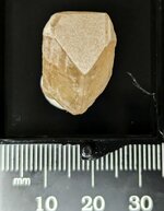
A piece of petrified wood, perhaps a small limb section or a twig.
This specimen is from the famous collecting site, Eden Valley, located near Eden, Sweetwater County, Wyoming. It is a rather bland looking example without the black core interior and the bluish gray associated chalcedony.
The first picture is in natural light and the example is of a near uniform tan coloration. The second picture is in LW 365nm and the specimen is a mottled orange color with hints of pink. The third picture is in MW 310nm and the specimen responds with a pastel orange. No response in SW 254nm.
This is the second specimen of fluorescent petrified wood to add to the Royal Collection. The other is a bright orange in LW and is from Texas.
Enjoy the photos!
Attachments
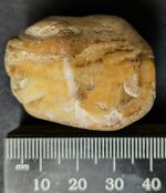
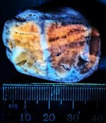
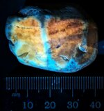
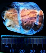
A specimen from western Germany.
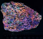
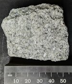
The rock consists of Nosean, a feldspathoid, intergrown with Sanadine, a high temperature K-feldspar. The specimen is from the East Eifel Volcanic Field (EEVF) within the Rhenish Massif, Rhineland-Palatinate State, Germany.
A classic Ed O’Dell gray to cream rock! HA HA The first picture is in natural light. The second picture is in LW 365nm, please note there are two fluorescent minerals present….orange nosean and orangish pink sanadine. Particularly the left 1/3rd of the image is the sanadine responding. The third picture is in MW 310nm and only the nosean responds orange, the sanadine has essentially no response. The fourth picture is in SW 254nm. The nosean is a strong orange and the sanadine is a distinctive red violet, as many K-feldspars typically display in SW.
KT finds it most interesting that the sanadine is responsive in both LW and SW, but not in MW!
Enjoy the pictures!
Attachments
-
 Nosean with sanadine, East Eifel Volcanic Field, Rhenish Massif, Rhineland-Palatinate State, W...jpg137.2 KB · Views: 204
 -
 Nosean with sanadine, East Eifel Volcanic Field, Rhenish Massif, Rhineland-Palatinate State, W...jpg116.7 KB · Views: 203
 -
 Nosean with sanadine, East Eifel Volcanic Field, Rhenish Massif, Rhineland-Palatinate State, W...jpg191.2 KB · Views: 213
 -
 Nosean with sanadine, East Eifel Volcanic Field, Rhenish Massif, Rhineland-Palatinate State, W...jpg169.5 KB · Views: 210
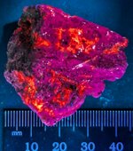
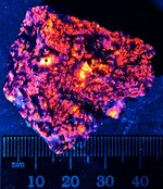
This morning KT got an email from a friend in northern Mississippi who suggested these pictures looked like different views looking into Hell!The rock consists of Nosean, a feldspathoid, intergrown with Sanadine, a high temperature K-feldspar. The specimen is from the East Eifel Volcanic Field (EEVF) within the Rhenish Massif, Rhineland-Palatinate State, Germany.
A classic Ed O’Dell gray to cream rock! HA HA The first picture is in natural light. The second picture is in LW 365nm, please note there are two fluorescent minerals present….orange nosean and orangish pink sanadine. Particularly the left 1/3rd of the image is the sanadine responding. The third picture is in MW 310nm and only the nosean responds orange, the sanadine has essentially no response. The fourth picture is in SW 254nm. The nosean is a strong orange and the sanadine is a distinctive red violet, as many K-feldspars typically display in SW.
Enjoy the pictures!
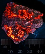
 When actually showing such specimens some people can hardly believe the said specimens are not hot, due to the glowing ember image, like associated with a camp fire.
A couple of nice specimens arrived today. One miniature which KT expected and a group of 2 gifts as thumbnails that are very interesting also!
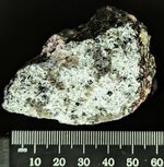
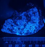
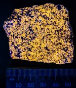
First to the miniature. The specimen consists of one of Ed’s gray and whitish rocks, from the Purple Passion Mine, Morgan Butte, Big Bug Mining District, Bradshaw Mountains, Yavapai County, Arizona. The minerals consist of fluorite, calcite, willemite and aragonite, all in shades of gray and cream as seen in picture 1.
But in picture 2, in LW 365nm, we begin to understand why the name of the mine! On this face of the specimen, there are two responsive minerals….fluorite is violet and aragonite is pale creamy white. In picture 3, in MW 310nm, we see that calcite makes its appearance as a bright red orange with a tiny bit of response by some selected grains of willemite with a pale green. Fluorite is still responsive as bluish violet. And in picture 4, taken in SW 254nm with 3 lamps,the fluorite is still responsive as blue, the calcite is a weaker orange, and the willemite is greenish as scattered grains throughout the fluorite matrix. The aragonite is unresponsive in both MW and SW.
Although only a miniature, this specimen is a mighty example of multi-colored fluorescence in different wavelengths of UV light!
Now to the thumbnails of tremolite in marble from the Nellie Ulmer Marble Quarry, Rockland, Knox County, Maine. One can see in the first picture a few of the lustrous fibrous crystal lathes of tremolite in the first picture, taken in natural light. But in picture 2, many more lathes of crystals are seen in the blue white response to SW254nm UV. KT imagines it would be a lot of fun to search for this mineral at night with a SW lamp!
Enjoy the pictures!
Attachments
-
 Fluorite, Calcite, Willemite, Aragonite, Purple Passion Mine, Morgan Butte, Big Bug Mining Dis...jpg220.2 KB · Views: 199
 -
 Fluorite, Calcite, Willemite, Aragonite, Purple Passion Mine, Morgan Butte, Big Bug Mining Dis...jpg154.1 KB · Views: 190
 -
 Fluorite, Calcite, Willemite, Aragonite, Purple Passion Mine, Morgan Butte, Big Bug Mining Dis...jpg125.9 KB · Views: 191
 -
 Fluorite, Calcite, Willemite, Aragonite, Purple Passion Mine, Morgan Butte, Big Bug Mining Dis...jpg156.5 KB · Views: 204
 -
 Tremolite xls, Nellie Ulmer Marble Quarry, Rockland, Knox Co., Maine Natural light.jpg113.8 KB · Views: 197
 -
 Tremolite xls, Nellie Ulmer Marble Quarry, Rockland, Knox Co., Maine SW 254nm.jpg97.1 KB · Views: 192
This sample arrived in the Post today!
A specimen that collectors call a quartz flower from a Dugway Geode, Dugway Geode Beds, Dugway Mountain, Juab County, Utah. The specimen was slightly larger, and KT trimmed off the non-fluorescent matrix that was not enhancing to the appearance of this piece, resulting in a nice miniature. The greenish fluorescence is attributed to traces of Uranium in the fluids that formed the quartz, being captured during formation.
This specimen is somewhat unusual in that it also fluoresces in LW 365nm and MW 310nm, as well as SW 254nm.
The first picture is in natural light, the second is in LW with the quartz displaying a grayish blue fluorescence. The third picture is in MW, and the quartz gives off a pastel green fluorescence. The 4th picture was taken in SW, and the mineral is a yellowish green.
Enjoy the photos!
Attachments
-
 Quartz flower, Dugway Geode Beds, Dugway Mtn., Juab Co., Utah, natural light.jpg95.4 KB · Views: 169
 -
 Quartz flower, Dugway Geode Beds, Dugway Mtn., Juab Co., Utah, LW 365nm.jpg140.6 KB · Views: 182
 -
 Quartz flower, Dugway Geode Beds, Dugway Mtn., Juab Co., Utah, MW 310nm.jpg83.3 KB · Views: 185
 -
 Quartz flower, Dugway Geode Beds, Dugway Mtn., Juab Co., Utah, SW 254nm.jpg85.8 KB · Views: 192
Long awaited packages arrived today!
Two packages containing 6 fluorescent specimens were a week and a half late in delivery from Pakistan, but it was not the mineral dealers fault, but some major fubars with the USPS, including it being necessary for KT to sign for them to receive them, and that had NEVER happened before on overseas packages!
Anyway, here is the info and pictures of the first 3 specimens…….
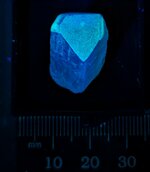
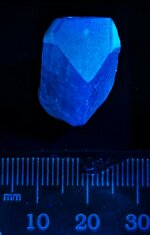
First specimen was supposed to be a terminated danburite from Badakhshan Province, Afghanistan. But it was neither terminated nor a danburite! It was a Scapolite miniature. Danburite fluoresces blue in LW, and this specimen fluoresces patchy orange in LW 365nm, pastel orange in MW 310nm, and a strong red in SW 254nm, as many scapolites do! So I have written the dealer, asking for my money back on this one specimen. I think it was likely an honest mistake, but at the least it should have been terminated! HA HA see the first 4 pictures. He just informed me that the danburite specimen shipped separately, but KT is still puzzled as to why He received a scapolite specimen He did not order! LOL
Pictures 5 and 6 are of a variety of Spodumene…..Kunzite from Kunar Province, Afghanistan, a nice hand specimen. Picture 5 is in natural light and Picture 6 is in LW 365nm, no response in either MW or SW. It is a nice cabinet specimen!
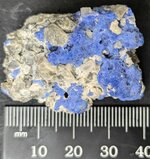
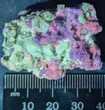
And pictures 7 and 8 are of Willemite on Albite with a few non-fluorescent black vesuvianite crystals (most on the reverse of the specimen) from Zagi, Khyber Pakhtankhwa Province, Pakistan. Picture 7 is in natural light and picture 8 is in SW 254nm, the Willemite responding the typical green, and the Albite a nice red.
Enjoy the pictures my friends!
Two packages containing 6 fluorescent specimens were a week and a half late in delivery from Pakistan, but it was not the mineral dealers fault, but some major fubars with the USPS, including it being necessary for KT to sign for them to receive them, and that had NEVER happened before on overseas packages!
Anyway, here is the info and pictures of the first 3 specimens…….
First specimen was supposed to be a terminated danburite from Badakhshan Province, Afghanistan. But it was neither terminated nor a danburite! It was a Scapolite miniature. Danburite fluoresces blue in LW, and this specimen fluoresces patchy orange in LW 365nm, pastel orange in MW 310nm, and a strong red in SW 254nm, as many scapolites do! So I have written the dealer, asking for my money back on this one specimen. I think it was likely an honest mistake, but at the least it should have been terminated! HA HA see the first 4 pictures. He just informed me that the danburite specimen shipped separately, but KT is still puzzled as to why He received a scapolite specimen He did not order! LOL
Pictures 5 and 6 are of a variety of Spodumene…..Kunzite from Kunar Province, Afghanistan, a nice hand specimen. Picture 5 is in natural light and Picture 6 is in LW 365nm, no response in either MW or SW. It is a nice cabinet specimen!
And pictures 7 and 8 are of Willemite on Albite with a few non-fluorescent black vesuvianite crystals (most on the reverse of the specimen) from Zagi, Khyber Pakhtankhwa Province, Pakistan. Picture 7 is in natural light and picture 8 is in SW 254nm, the Willemite responding the typical green, and the Albite a nice red.
Enjoy the pictures my friends!
Attachments
-
 Scapolite, Badakhshan, Afgh., natural light.jpg174.9 KB · Views: 181
 -
 Scapolite, Badakhshan, Afgh., LW 265nm.jpg106.8 KB · Views: 193
 -
 Scapolite, Badakhshan, Afgh., MW 310nm.jpg99.7 KB · Views: 177
 -
 Scapolite, Badakhshan, Afgh., SW 254nm.jpg97 KB · Views: 197
 -
 Kunzite, Kunar Province, Afghanistan, natural light.jpg147.7 KB · Views: 193
 -
 Kunzite, Kunar Province, Afghanistan, LW 365nm.jpg186.7 KB · Views: 180
 -
 Willemite on Albite, Zagi, KPK, Pakistan, natural light.jpg152.4 KB · Views: 181
 -
 Willemite on Albite, Zagi, KPK, Pakistan, SW 254nm.jpg110.6 KB · Views: 193
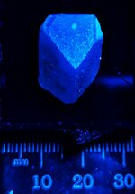
A package arrived today, and KT is pleased to announce that the actual real Danburite arrived safe and sound. My Royal confusion over this situation is due to two things happening. First, eBay announced that this specimen arrived yesterday, but what I got in with the other 5 specimens is that Scapolite that I had not ordered, apparently a mistake or perhaps a gift from the first eBay dealer. There was no label, so this was quite confusing to me. However, the real dealer sent me a message saying it should arrive tomorrow and to have patience….HA HA. And it did arrive! Today!
This specimen, a single T/N crystal of Danburite is from Kokcha Valley,Badakhshan Province, Afghanistan. Thanks to Mindat.org for a bit more precise location, at least as to the Valley. There are 15 collecting sites within that valley so that is as close as KT can get.
Anyway,the first picture is in natural light, the specimen mounted in a Perky Box. The second picture is in LW 365nm, with a nice blue response. The third picture is in MW 310nm, also with a blue response, and the fourth picture is in SW 254nm, again with a blue response! A bit unusual to have the same response across all wavelengths of UV.
Enjoy the photos!
Attachments
-
 Danburite, Kokcha Valley, Badakhshan Province, Afghanistan, natural light.jpg98.9 KB · Views: 189
 -
 Danburite, Kokcha Valley, Badakhshan Province, Afghanistan, LW 365nm.jpg62 KB · Views: 188
 -
 Danburite, Kokcha Valley, Badakhshan Province, Afghanistan, MW 310nm.jpg53.2 KB · Views: 189
 -
 Danburite, Kokcha Valley, Badakhshan Province, Afghanistan, SW 254nm.jpg53 KB · Views: 183
Here are the last 3 specimens that arrived yesterday in those two packages….all of these were expected samples.
The first is a miniature of blue Sodalite, var. hackmanite, from Badakhshan Province, Afghanistan. The first picture is in natural light, and the second picture is in LW 365nm….little to no response in either MW or SW.
The second specimen is a crystal of Fluorapatite perched on a mica book on feldspar from Pakistan. The first picture of it is in natural light, the second pic is in LW 365nm, the third one is in MW 310nm,and the fourth one is in SW 254nm. Very strongly fluorescent crystal!
The third specimen consists of Scapolite, var. marialite, in calcite matrix. The first picture is in natural light, the second is in LW365nm, the third is in MW 310nm (note the calcite matrix displaying red fluorescence), and the fourth picture is in SW 254nm, with the calcite also nicely displaying red. The calcite matrix exhibits a distinct and fairly long term red phosphorescence after exposure to SW UV.
Enjoy the pictures!
Attachments
-
 Sodalite, var. hackmanite, Badakhshan Province, Afghanistan, natural light.jpg195 KB · Views: 187
 -
 Sodalite, var. hackmanite, Badakhshan Province, Afghanistan, LW 365nm.jpg197.2 KB · Views: 185
 -
 Fluorapatite on mica and feldspar, Pakistan, natural light.jpg147.8 KB · Views: 183
 -
 Fluorapatite on mica and feldspar, Pakistan, LW 365nm.jpg122.7 KB · Views: 196
 -
 Fluorapatite on mica and feldspar, Pakistan, MW 310nm.jpg102.8 KB · Views: 181
 -
 Fluorapatite on mica and feldspar, Pakistan, SW 254nm.jpg85 KB · Views: 186
 -
 Scapolite, var. marialite, in calcite matrix, Afghanistan, natural light.jpg168.6 KB · Views: 185
 -
 Scapolite, var. marialite, in calcite matrix, Afghanistan, LW 365nm.jpg129 KB · Views: 182
 -
 Scapolite, var. marialite, in calcite matrix, Afghanistan, MW 310nm.jpg111.5 KB · Views: 191
 -
 Scapolite, var. marialite, in calcite matrix, Afghanistan, SW 254nm.jpg140.1 KB · Views: 189
Some early Christmas arrivals, thanks to the USPO!
The first 4 pictures are of a Chert pebble from the shoreline of Lake Michigan at Fox Point in Milwaukee County, Wisconsin. It was listed as being fluorescent in LW UV, however much to my surprise it is fluorescent in LW, MW, and SW!
The first picture is in natural light. The second picture is in LW365nm, the third picture is in MW 310nm, and the fourth picture is in SW 254nm. The seller stated that he had no idea what caused the fluorescence, and he speculated the presence of calcite. But KT does not think so, it likely contains a bit of organic matter, and just as in many petrified wood specimens, that is what causes the orange fluorescence. Just a thought.
The second set of pictures are of Scheelite ore from east of Tucson in Cochise County, Arizona. Unfortunately that is as close as KT can get on a location as Mindat.org lists 130 localities in that county as having Scheelite. His Majesty does know that it is from a desert environment as it has caliche on the backside of the specimen, caliche being formed in soil and as coatings on rocks in arid environments. The Scheelite was listed as being fluorescent in SW UV, but in this specimen it is also fluorescent in LW, MW, and SW.
The first picture is in natural light. The second picture is in LW365nm, the third picture was taken in MW 310nm, and the last picture was taken in SW 254nm.
Enjoy the pictures, and Merry Christmas to all!
Attachments
-
 Chert pebble from Lake Michigan shoreline at Fox Point, Milwalkee Co., Wisconsin, natural light.jpg115.3 KB · Views: 181
 -
 Chert pebble from Lake Michigan shoreline at Fox Point, Milwalkee Co., Wisconsin, LW 365nm.jpg135.9 KB · Views: 178
 -
 Chert pebble from Lake Michigan shoreline at Fox Point, Milwalkee Co., Wisconsin, MW 310nm.jpg96.9 KB · Views: 176
 -
 Chert pebble from Lake Michigan shoreline at Fox Point, Milwalkee Co., Wisconsin, SW 254nm.jpg135.1 KB · Views: 168
 -
 Scheelite ore, east of Tucson, in Cochise Co., AZ, natural light.jpg219.8 KB · Views: 173
 -
 Scheelite ore, east of Tucson, in Cochise Co., AZ, LW 365nm.jpg118.5 KB · Views: 177
 -
 Scheelite ore, east of Tucson, in Cochise Co., AZ, MW 310nm.jpg142.6 KB · Views: 192
 -
 Scheelite ore, east of Tucson, in Cochise Co., AZ, SW 254nm.jpg144.4 KB · Views: 196
Two fluorescent specimens arrived today in the Royal Mailbox! Both are from overseas.
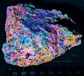
The first specimen consists of 2 Zircon crystal T/Ns from the Chilwa Alkaline Province, Mount Malosa area, Rift Mineral Province, Malawi, Africa. The first picture is in natural light, the second is in MW 310nm UV and the third is in SW 254nm UV. There was very weak and dull orange fluorescence in LW 365nm, but KT decided not to try and take an image of it. If you investigate Zircons from Malawi, Africa on Mindat.org, you will find about 40 something localities in southern Malawi. Looking over the 80 something pictures, only one general locality or area is producing zircons of this size and color with this fluorescence in abundance. Within that general location there are about 12 sites, some unnamed and poorly documented so despite the label looking rather specific, it is a general area of where these crystals came from!
The second specimen is green Hauyne on a marble matrix from Sar-e-Sang, Kuran wa Munjan District, Badakhshan Province, Afghanistan. Sar-e-Sang is a valley containing about 15 sites, some yielding gem Sodalite and gem Afghanite. When I reviewed the pictures of Hauyne in Mindat.org, there were only 2 pictures of green Hauyne, both of the same specimen and its locality is what KT listed, nothing more specific. To see this mineral as green instead of intense blue is pretty unusual. The first picture is in natural light, and the Hauyne is green with a bluish cast, this being a fairly rare color. The second picture is in LW 365nm and the third picture is in MW 310nm. No response in SW 254nm.
Enjoy the pictures!
Attachments
-
 Zircon xls, Chilwa Alk. Prov., Mount Amlosa area, Rift Min. Prov., Malawi, Africa, natural light.jpg116.2 KB · Views: 181
 -
 Zircon xls, Chilwa Alk. Prov., Mount Amlosa area, Rift Min. Prov., Malawi, Africa, MW 310nm.jpg100.3 KB · Views: 174
 -
 Zircon xls, Chilwa Alk. Prov., Mount Amlosa area, Rift Min. Prov., Malawi, Africa, SW 254nm.jpg128.6 KB · Views: 174
 -
 Hauyne on marble, Sar-e-Sang, Kuran wa Munjan District, Badakhshan Prov., Afghanistan, natural...jpg260.7 KB · Views: 182
 -
 Hauyne on marble, Sar-e-Sang, Kuran wa Munjan District, Badakhshan Prov., Afghanistan, LW 365nm.jpg198.1 KB · Views: 179
 -
 Hauyne on marble, Sar-e-Sang, Kuran wa Munjan District, Badakhshan Prov., Afghanistan, MW 310nm.jpg156.2 KB · Views: 184
Another specimen arrived in the Royal Mailbox today! First of 2025!
A nice miniature of Wollastonite from the well known Sterling Hill Mine, Ogdensburg, Sussex County, New Jersey. The seller had informed KT that the specimen looked better under his SW lamp than the pictures he was able to take.
The first picture is in natural light, and the rock has a uniform crystalline texture, one might say it is holocrystalline. Wollastonite typically forms as elongate crystals, in this specimen they have a ratio of about 1:2, width to length, and the mineral composes most of the rock specimen, so the rock is equigranular overall. A nice sized miniature.
The second picture is in SW 254nm and the Wollastonite responds with an orangish yellow. The original photograph looked too yellow when compared to the actual view with the naked eye, so KT made minor adjustments to the color of the image to look like the raw specimen, both in tint and intensity. No response in either LW or MW UV.
Enjoy the pictures!
Attachments
KT got bored so this post is the result!
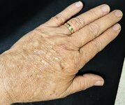
When giving UV presentations to groups, KT often shows the participants the back of his Royal hand, first in natural light and then in LW 365nm, to demonstrate to people how long term exposure to the sun can damage your skin. At 75 years old, the Royal skin is certainly displaying the long term effects of sunbathing when a teenager, and working outdoors without using any protective lotions. Fortunately His Majesty has had no skin cancers or melanomas show up, but He does have a few flat moles and skin tags that are inspected regularly just to see that they are not suddenly growing or changing shapes or darkening.
Anyway, His Majesty is a child of the 50s, and my parents had no knowledge of what the sun could do, other than give you a sunburn if you stayed out in it too long.
So there is always something interesting to come across with UV lights! HA HA
Check out the two pictures below! Note the loss of pigmentation in the LW image and the large number of wrinkles, not visible in natural light. Once you view the loss of pigment in UV you can readily see it also in natural light.
Attachments

A small arrival in the Royal Mail Box today!
This is a variety of Quartz, Chalcedony, that locally is called a Chalcedony rose, from just south of Deming, Luna County, New Mexico.
It displays very weak pale greenish fluorescence in LW 365nm, but did not display that color in the LW photos, so none are shown. The eye is so much more sensitive that a camera photocell! HA HA Anyway, this is a small, but complete specimen, no bigger than His Majesty's pinky-finger fingernail, as you can tell from the mm scale.
The first picture is in natural light, and the specimen is slightly pink and milky and displays formational banding. The second picture isin MW 310nm and displays a reasonable yellowish green. The third picture is in SW 254nm and the specimen displays a strong yellowish green response. The green fluorescence is typical of minerals that contain a trace of Uranium.
The seller stated it was a “cave formation”, and it might be, but KT seriously doubts it, tho it obviously formed within a pocket in the host rock, perhaps even in the central cavity of some of the local agates that are well documented in this county.
Enjoy the pics.
Attachments
Today's Royal Mailbox Specimen!
This is an encrusting example of Hyaline, Opal-An, from Old 20 Mine, Crabtree Creek, Spruce Pine District, Yancey County, North Carolina. The specimen is a miniature. The first picture was taken in natural light, the second in LW 365nm, the third in MW 310nm, and the last one is in SW254nm. All fluorescent green, and the green gets stronger as one ranges from 365nm to SW 254nm, but is nice in any UV light.
This is the characteristic response of Hyaline when it contains a trace of Uranium!
Enjoy the pictures!
Attachments
-
 Hyaline, Opal-An, Old 20 Mine, Crabtree Creek, Spruce Pine District, Burnsville, Yancey Co., N...jpg222.9 KB · Views: 162
 -
 Hyaline, Opal-An, Old 20 Mine, Crabtree Creek, Spruce Pine District, Burnsville, Yancey Co., N...jpg220.7 KB · Views: 168
 -
 Hyaline, Opal-An, Old 20 Mine, Crabtree Creek, Spruce Pine District, Burnsville, Yancey Co., N...jpg115.1 KB · Views: 174
 -
 Hyaline, Opal-An, Old 20 Mine, Crabtree Creek, Spruce Pine District, Burnsville, Yancey Co., N...jpg194.9 KB · Views: 166
Odds and Ends
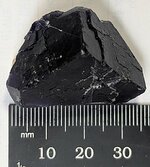
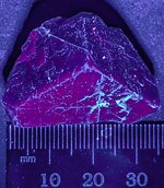
This cleaved crystal of fluorite is from China. KT had hoped to get a more specific location, but has had no luck. One can find these specimens on eBay, listed under Fluorescent Black Rose Fluorite, marketed by both Chinese and Pakistani dealers. But no one locates it closer than China. There is one place in Mexico that produces this oddly fluorescent fluorite, but the mineral is not heat treated,but essentially all of it from China is heat treated, which changes the typical blue response to a pale to bright to dark red fluorescence. The first picture is in natural light, and the second in LW 365nm. Sometimes on the back side of these specimens, or on cleavages, the red fluorescence shows up as banding that follows zoning in the crystal, likely due to some specific trace element that was incorporated from the formational fluids during growth.
The other unique thing is that the red shows up more dramatically, like a flash and then fades, when the LW UV hits it. Unfortunately KT could not show that. The red is much stronger to the naked eye that it is in the image, due to the differences between the human eye and the photocell of the camera.
Enjoy the pics.
Attachments
Similar threads
- Replies
- 8
- Views
- 130