An interesting Thumbnail specimen arrived today in the Royal Mailbox!
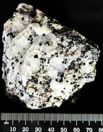
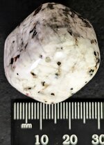
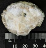
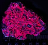
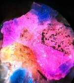
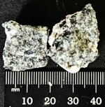
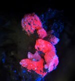
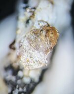
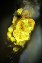
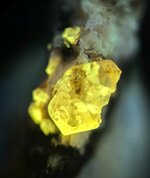
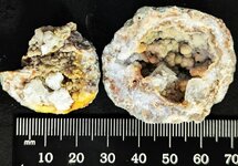
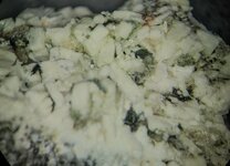
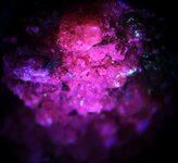
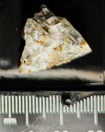
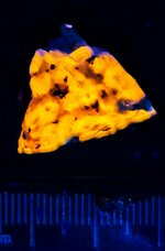
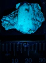
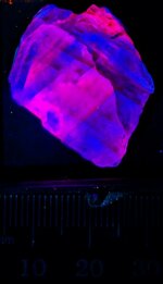
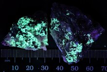
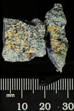
It is a single Fluorapatite crystal from Imilchil, Imilchil Cercele, Midelt Province, Morocco, nicely double terminated. There are many of these crystals photographed from this locality in Mindat.org.
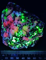
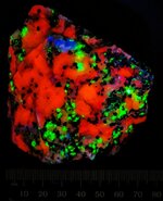
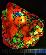
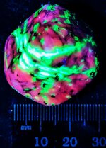
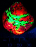
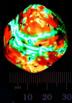
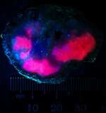
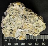
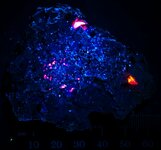
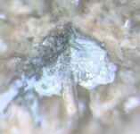
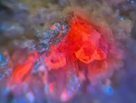
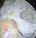
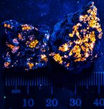
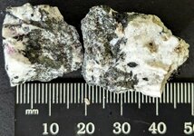
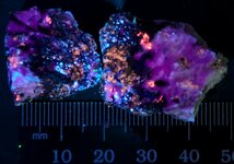
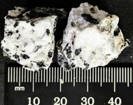
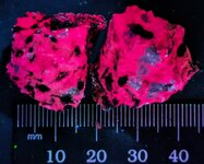
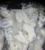
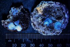
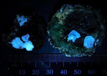
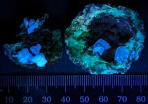
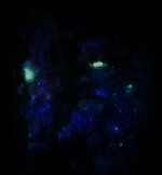
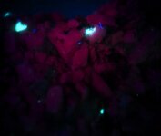
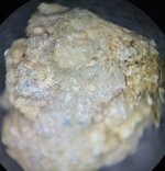
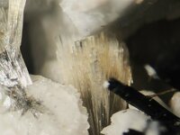
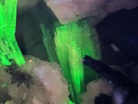
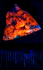
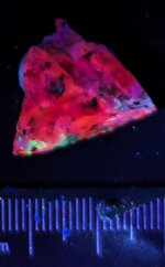
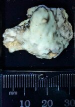
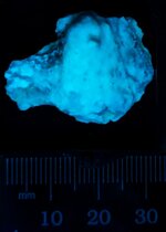
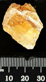
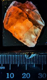
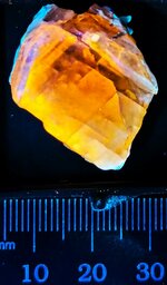
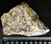
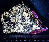
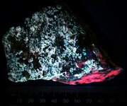
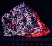
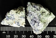
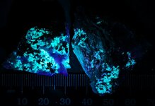
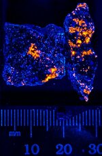
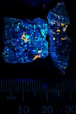
It is from the Sobolewski Collection. Unfortunately, KT does not know who this gentleman was. The first picture is in natural light, the second in LW 365nm, the third in MW 310nm, and the fourth, and last one, is in SW254nm. Interestingly different response in each distinct wavelength.
Anyway, it was remounted in a Perky box (1.25" cube) and will soon be filed away in KT's new Royal M/M and T/N cabinet, in drawer 3 with some others!
Enjoy the pictures!
It is a single Fluorapatite crystal from Imilchil, Imilchil Cercele, Midelt Province, Morocco, nicely double terminated. There are many of these crystals photographed from this locality in Mindat.org.
It is from the Sobolewski Collection. Unfortunately, KT does not know who this gentleman was. The first picture is in natural light, the second in LW 365nm, the third in MW 310nm, and the fourth, and last one, is in SW254nm. Interestingly different response in each distinct wavelength.
Anyway, it was remounted in a Perky box (1.25" cube) and will soon be filed away in KT's new Royal M/M and T/N cabinet, in drawer 3 with some others!
Enjoy the pictures!
Attachments
-
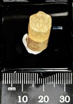 Fluorapatite, double terminated, Imilchi, Imilchi Cercela, Midelt Province, Morocco, natural l...jpg89.3 KB · Views: 188
 -
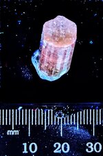 Fluorapatite, double terminated, Imilchi, Imilchi Cercela, Midelt Province, Morocco, LW 365nm.jpg101.4 KB · Views: 181
 -
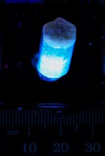 Fluorapatite, double terminated, Imilchi, Imilchi Cercela, Midelt Province, Morocco, MW 310nm.jpg35.5 KB · Views: 184
 -
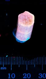 Fluorapatite, double terminated, Imilchi, Imilchi Cercela, Midelt Province, Morocco, SW 254nm.jpg40.1 KB · Views: 190