Now to some Arkansas fluorescent specimens.
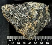
These two specimens were collected from the lower level of the main pit of the Martin Marietta Quarry during the 5th Annual CUSMS gathering on the Wednesday, October 11, 2023 field trip by His Majesty, thanks to Ed O’Dell bringing it to my Royal attention! The host rock consists of contact metamorphosed shale from the Stanley Formation (Mississippian). The mineralization was first thought to consist of only fluorite in a thin veinlet, less than 2 mm, that had fractured in the plane of the vein during blasting. Further investigations with a LW 365nm lamp proved otherwise for one of the specimens!
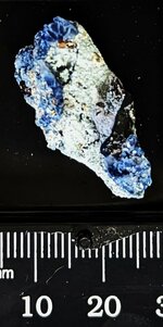
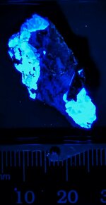
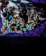
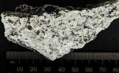
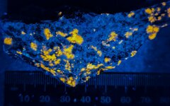
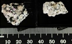
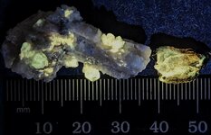
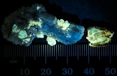
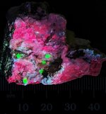
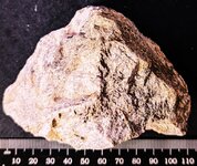
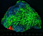
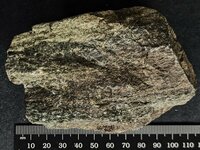
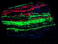
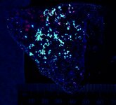
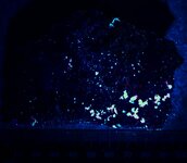
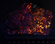
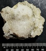
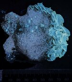
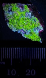
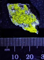
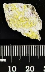
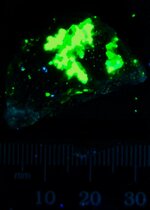
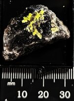
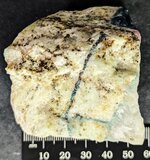
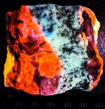
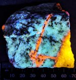
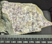
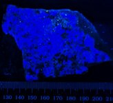
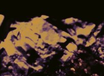
The first piece is a small cabinet specimen of a fluorite-calcite veinlet, well exposed on a fracture surface. Pieces of this mineralization were scattered about on the northwest end of the quarry floor. Several examples were collected for UV examination later. This specimen is the only one that had both fluorescent fluorite and calcite in the veinlet. The first picture is in natural light and the second is in LW 365nm, the fluorite displaying its typical blue coloration and the calcite displaying orange.
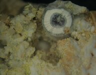
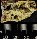
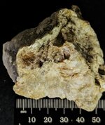
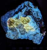
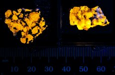
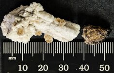
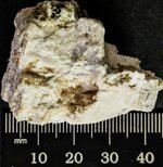
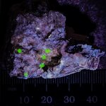
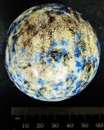
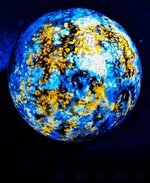
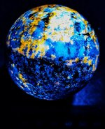
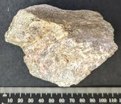
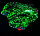
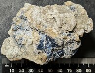
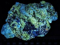
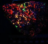
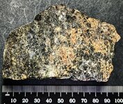
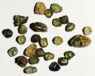
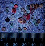
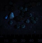
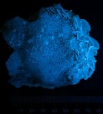
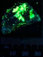
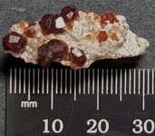
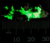
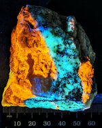
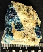
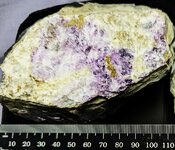
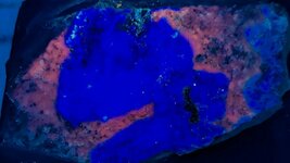
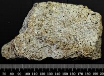
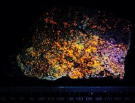
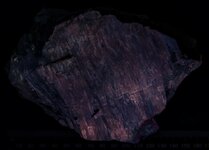
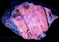
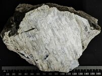
The second specimen just shows the general appearance of the broken veinlet surface, and looks like perhaps there are other minerals present in the natural light image (Picture 1), but Picture 2 shows a strong fluorite response to LW 365nm and enough coverage to assume that some of the fluorite is colorless, some purple, and some white from the uniform blue coloration.
Enjoy the pictures. Several more to come from these field trips!
These two specimens were collected from the lower level of the main pit of the Martin Marietta Quarry during the 5th Annual CUSMS gathering on the Wednesday, October 11, 2023 field trip by His Majesty, thanks to Ed O’Dell bringing it to my Royal attention! The host rock consists of contact metamorphosed shale from the Stanley Formation (Mississippian). The mineralization was first thought to consist of only fluorite in a thin veinlet, less than 2 mm, that had fractured in the plane of the vein during blasting. Further investigations with a LW 365nm lamp proved otherwise for one of the specimens!
The first piece is a small cabinet specimen of a fluorite-calcite veinlet, well exposed on a fracture surface. Pieces of this mineralization were scattered about on the northwest end of the quarry floor. Several examples were collected for UV examination later. This specimen is the only one that had both fluorescent fluorite and calcite in the veinlet. The first picture is in natural light and the second is in LW 365nm, the fluorite displaying its typical blue coloration and the calcite displaying orange.
The second specimen just shows the general appearance of the broken veinlet surface, and looks like perhaps there are other minerals present in the natural light image (Picture 1), but Picture 2 shows a strong fluorite response to LW 365nm and enough coverage to assume that some of the fluorite is colorless, some purple, and some white from the uniform blue coloration.
Enjoy the pictures. Several more to come from these field trips!
Attachments
-
 Fluorite and Calcite, Martin Marietta Quarry, Hot Spring Co., AR, natural light.jpg190.7 KB · Views: 136
 -
 Fluorite and Calcite, Martin Marietta Quarry, Hot Spring Co., AR, LW 365nm.jpg67.3 KB · Views: 155
 -
 Fluorite, Martin Marietta Quarry, Hot Spring Co., AR, natural light.jpg173.3 KB · Views: 135
 -
 Fluorite, Martin Marietta Quarry, Hot Spring Co., AR, LW 365nm.jpg73.7 KB · Views: 156
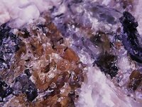
 It is from the California State Gem Mine, San Benito Co., California, and that is the type locality for both these minerals. The Joaquinite-(Ce) is a micro crystal and not fluorescent, but the much more abundant Benitoite is of course fluorescent blue white in SW 254nm. The German label read: Benitoit, Kalifornien, USA!
  KT remounted it in a Perky 1.25" cube box because the base is black, non-fluorescing plastic, so a better picture.