Was a wait but it was worth it!
Finally today, a couple more interesting fluorescent minerals arrived in the Royal Mail Box and it has surely taken KT out of the Covid malaise a bit!
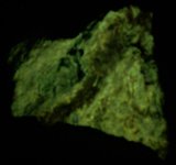
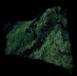
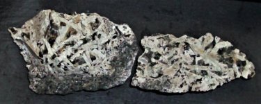
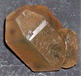
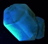
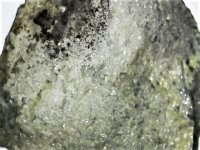
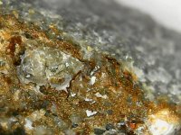
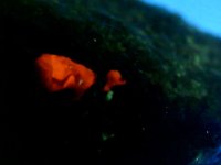
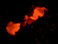
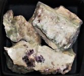
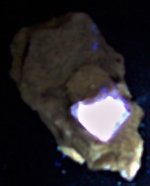
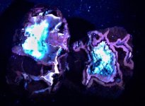
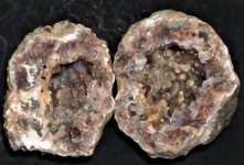
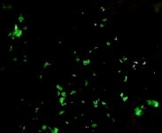
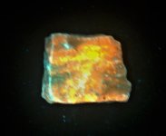
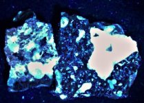
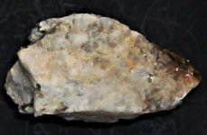
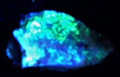
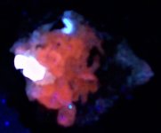
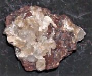
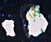
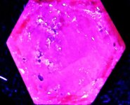
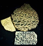
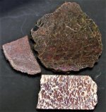
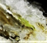
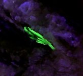
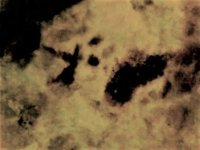
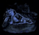
The first pair of pictures are of Montbrasite from Emmons Quarry, near Greenwood, Oxford County, Maine. The specimen is a miniature and shown in the first picture in natural light, and in the second picture is shown in SWUV 254nm light...a nice change!
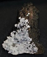
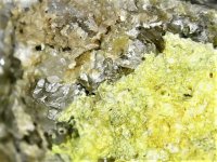
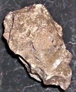
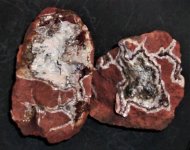
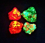
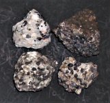
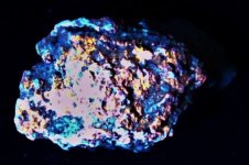
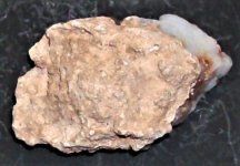
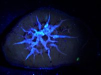
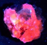
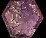
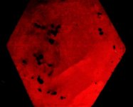
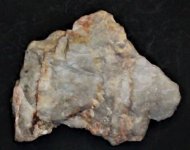
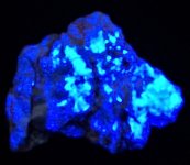
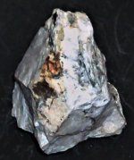
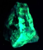
The second specimen was sold as Calamine (the old name for hemimorphite) and listed from Searey County, Arkansas, which was a misspelling of the county name which actually is Searcy County. Anyway, in his former collecting days, KT visited many of the old zinc mines in north Arkansas, including those 10 or so in Searcy County. But alas, His Majesty was not interested in fluorescent minerals at the time and never checked any of the samples He collected.
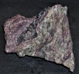
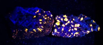
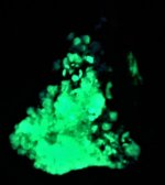
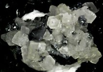
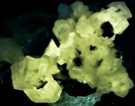
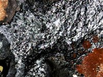
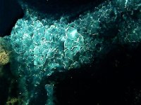
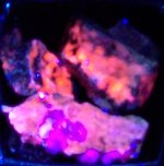
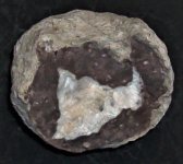
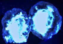
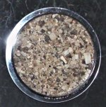
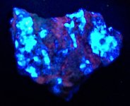
Anyway, when the specimen arrived, KT was suspicious about the identification...as it was accompanied by an old New Hampshire University Collection label. Now Hemimorphite is a zinc silicate, and smithsonite is a zinc carbonate. Both have been identified from those mines, but every specimen of hemi KT has ever seen or collected from Arkansas was crystalline and the crystals were somewhat sword blade in form, whereas smithsonite is commonly mamillary in form, as this specimen. So KT took a tiny piece of the fluorescing mineral and put a drop of acid on it and observed under his binocular microscope. Lots of fizzing and bubbling going on! So it is smithsonite and not calamine. No telling why the museum staff never tested it! Anyway, the first picture is in natural light. Specimen is hand sized, and the second picture is in LWUV 365nm.
Enjoy the pictures!
Finally today, a couple more interesting fluorescent minerals arrived in the Royal Mail Box and it has surely taken KT out of the Covid malaise a bit!
The first pair of pictures are of Montbrasite from Emmons Quarry, near Greenwood, Oxford County, Maine. The specimen is a miniature and shown in the first picture in natural light, and in the second picture is shown in SWUV 254nm light...a nice change!
The second specimen was sold as Calamine (the old name for hemimorphite) and listed from Searey County, Arkansas, which was a misspelling of the county name which actually is Searcy County. Anyway, in his former collecting days, KT visited many of the old zinc mines in north Arkansas, including those 10 or so in Searcy County. But alas, His Majesty was not interested in fluorescent minerals at the time and never checked any of the samples He collected.
Anyway, when the specimen arrived, KT was suspicious about the identification...as it was accompanied by an old New Hampshire University Collection label. Now Hemimorphite is a zinc silicate, and smithsonite is a zinc carbonate. Both have been identified from those mines, but every specimen of hemi KT has ever seen or collected from Arkansas was crystalline and the crystals were somewhat sword blade in form, whereas smithsonite is commonly mamillary in form, as this specimen. So KT took a tiny piece of the fluorescing mineral and put a drop of acid on it and observed under his binocular microscope. Lots of fizzing and bubbling going on! So it is smithsonite and not calamine. No telling why the museum staff never tested it! Anyway, the first picture is in natural light. Specimen is hand sized, and the second picture is in LWUV 365nm.
Enjoy the pictures!
Attachments
-
 Montbrasite, Emmons Quarry, Oxford Co., MA miniature, natural light.JPG121.4 KB · Views: 110
 -
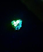
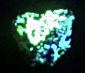
 Montbrasite, Emmons Quarry, Oxford Co., MA miniature, SWUV 254nm.JPG61.3 KB · Views: 115
 -
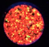
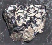
 Smithsonite, north Searcy Co., AR, hand specimen, natural light.JPG139.9 KB · Views: 113
 -
 Smithsonite, north Searcy Co., AR, hand specimen, LWUV 365nm.JPG59.1 KB · Views: 110